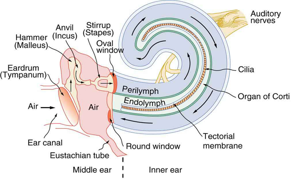
The organ of Corti contains hair cells that respond to vibration by brushing their stereocilia against a fixed structure called the tectorial membrane. The basilar membrane contains a specialized structure known as the organ of Corti that plays a key role in auditory transduction. The structure separating the scala tympani from the cochlear duct is known as the basilar membrane. The Reissner membrane separates the scala vestibuli from the cochlear duct and the stria vascularis, specialized cells lining the lateral wall of the cochlear duct.

The Reissner membrane maintains the differences in ion concentration between the endolymph and perilymph. This positive potential results from high potassium and low sodium ions concentrations compared to the surrounding perilymph. This hollow, bony tube contains endolymph, which has a higher positive potential than the surrounding perilymph. The cochlear duct lies between the scala vestibuli and the scala tympani. Īt the end of the scala tympani, the vibrations within the perilymph displace the round window. The vibrations then travel up the cochlea to the apex through a hollow bony tube called the scala vestibuli and then from the apex to the base of the cochlea through another hollow, bony tube called the scala tympani. The footplate of the stapes contacts the oval window in a piston-like motion that transmits vibration to perilymph within the cochlea. These vibrations are then transferred to the middle ear along the ossicular chain of the malleus, incus, and stapes. The process of auditory transduction begins with sound waves entering the external acoustic meatus and striking the tympanic membrane, resulting in vibration. These electrical impulses travel to the brain via the vestibulocochlear nerve (CN VIII). Sound waves cause vibrations, which bend the hair cell stereocilia via an electromechanical force, leading to electrical impulses. There is one single row of inner hair cells (IHC) and three rows of outer hair cells (OHC), separated by the tunnel of Corti. These hair cells are topped with hair-like structures called stereocilia. As previously mentioned, these sensory hair cells are activated by the potential difference between the perilymph and the endolymph. The organ of Corti is a cellular layer sitting on top of the BM in which sensory hair cells are found. The osseous spiral lamina and BM separate the SM from the ST, with the BM determining the mechanical wave propagation properties. The SL contains blood vessels and fibrocytes, which play an important role in immune response and ion homeostasis. The spiral ligament (SL), connected to the Reissner membrane and the basilar membrane (BM), forms the outer wall of the scala media. The Reissner membrane is what separates the SV from the SM. The helicotrema is the apex of the cochlea and is where the ST and SV meet. The stria vascularis is highly vascularized tissue that lines the lateral walls of the cochlea and is responsible for producing the endolymph for the SM and maintaining the ion balance surrounding the outer and inner hair cells of the organ of Corti. This electrical potential difference allows potassium ions to flow into the hair cells during mechanical stimulation of the hair bundle-discussed later in further detail. As there is a higher potassium concentration than sodium, the endolymph's electrical potential is approximately 80-90 mV more positive than perilymph. The composition of endolymph resembles that of intracellular fluid, with potassium as the primary cation. The perilymph contains proteins, such as enzymes and immunoglobulins, which are important for metabolism and the immune response. The composition of perilymph resembles that of extracellular fluid, with sodium as the primary cation, and, via the perilymphatic duct, serves as a connection to the CSF of the subarachnoid space. The SV and ST are filled with perilymph, while the SM is filled with endolymph.

The cochlear tube is formed by three membranous and fluid-filled chambers that run parallel to each other the scala vestibuli (SV) or vestibular duct, the scala media (SM) or cochlear duct, and the scala tympani (ST) or tympanic duct. The bony labyrinth is a cavity within the temporal bone consisting of the vestibule, semicircular canals, and cochlea. The inner ear is comprised of a bony and membranous labyrinth. Understanding cochlear anatomy is essential to understanding its physiology.


 0 kommentar(er)
